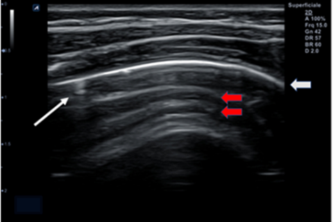
Introduction COVID-19 in newborns is a variable disease that remains relatively understudied. In symptomatic newborns, infection may progress with subtle or non-specific clinical and laboratory findings. Lung ultrasound (LUS) can serve as a valuable imaging tool for evaluating lung damage caused by COVID-19, all without radiation exposure. Additionally, the long-term effects of COVID-19 in the neonatal population are still inadequately investigated. Population and Methods A retrospective cohort study was conducted from January to December 2022 at the Neonatal Intensive Care Unit (NICU) of Monaldi Hospital in Naples, which serves as a hub for managing newborns with COVID-19 in our region. The diagnosis of SARS-CoV-2 infection was established through the analysis of nasal/oral-pharyngeal swabs using RT-PCR molecular methods. Clinical, laboratory, and instrumental parameters of each newborn were assessed during hospitalization and a 6-month follow-up period. LUS was performed in the supine, prone, and lateral positions, with the results compared to Chest-X-ray (CXR). Results A total of 31 patients with positive RT-PCR results were admitted. Of these, 65% were male, with a median weight of 3514.1 ± 560.2 grams and a median corrected age of 39 ± 2 weeks. The median age at admission was 21.1 ± 12.2 days. The median time for naso-pharyngeal swab negativization was 13.3 ± 5.4 days. The most common clinical manifestations in our sample included fever (64.5%), respiratory distress (16%), and gastrointestinal symptoms (10%). Antibiotics for suspected superimposed bacterial infections were administered in 16% of cases, and hemodynamic support was required in 6%. Additionally, 16% of infants needed respiratory support, either in the form of high-flow nasal cannula (HHHFNC) or oxygen therapy. Pathological findings were observed in approximately 10% of infants on chest X-rays, while lung ultrasound (LUS) revealed alterations in 22.5% of cases. The predominant LUS pattern identified was characterized by A-lines (horizontal reverberation artifacts), with a few B-lines (ring-down artifacts), and a thin pleural hyperechoic line. Correlation analysis showed that the time for swab negativization was not influenced by other variables. Higher ferritin values at admission were associated with a more severe disease course, particularly in terms of respiratory distress and the need for ventilatory support (ρ 0.58; p 0.001). The ROC curve indicated 82% sensitivity and 58% specificity for this parameter. During the 6-month follow-up, all infants exhibited regular growth, a healthy appearance, and normal vital signs. Mild to moderate transient neutropenia was observed in 25.8% of infants. Only one infant displayed persistent LUS alterations (a few B-lines) at 1 and 3-month follow-up, which were no longer visible at the 6-month evaluation. Conclusion The overall data from our population indicated favorable outcomes in infants affected by COVID-19. Higher ferritin values at admission were associated with a more severe disease course. The most common LUS pattern observed included a reduction in A-lines, an increase in/fusion of B-lines, and, at times, an abnormal/fragmented hyperechoic pleural line. LUS appeared to be a sensitive and non-invasive tool for the comprehensive evaluation of pulmonary pathology caused by COVID-19 in newborns.