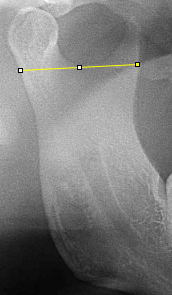
Age estimation using Orthopantomographs – a study on radiomorphometric parameters of mandibular ramus and gonial angle in south Indian population.Abhishek Gupta,1 Vijayalaxmi K R,21 Oral Medicine and Radiology, Chitwan Medical College, Chitwan, 44207, Nepal.2 Oral Medicine and Radiology, Government Dental College and Research Institute, Bangalore,560001, India.Correspondence should be addressed to Abhishek Gupta; [email protected] . Age estimation using orthopantomographs (OPGs) has been in practice for a long time, but none of the methods is considered universally accepted yet. Very few studies have been conducted using radiomorphometric indices for age estimation. This study was conducted to assess the radiomorphometric indices using digital OPGs and their applicability for age estimation of individuals in south Indian population. Materials and Methods . OPGs of 611 individuals ( 302 male and 309 females) were selected on basis of predetermined inclusion and exclusion criteria. The OPGs of the patients ( age 0 to 69 years) were divided into 7 groups ( each decade) . The 6 radiomorphometric indices of bilateral Mandible ( upper and lower Ramus breadth; condylar , projected and coronoid ramus height ; and gonial angle ) were measured on Monitor of 15” display LED Screen using IMAGE J software.Results. The mean gonial angle was found to be higher in females as compared to males in all the age groups (with maximum in 10-19years age groups ) except in 03-09years age groups. In contrast to that, mean condylar height , mean projected condylar height , mean coronoid ramus height, and mean ramus breadth were found to be higher in males as compared to females in all the age groups except in 03-09 years groups, which was statically significant. Gonial angle was found to be greater in 3 to 19years age group, and thereafter decreasing with age. Condylar height , projective condylar height and coronoid ramus height were found to be increasing during 2nd and 3rddecades i.e 19 to 39 age group and thereafter decreasing with age (positive correlation ). Conclusion . Condylar height was found to be the most significant age predictor and ramus breadth was found to be most insignificant age predictor.IntroductionAge assessment involves many factors with limited precision.1 One of the simple and accurate maturational indicator is skeletal development.2 Age bring changes in teeth and bone including the facial bones.3 Also, these structures remains for a very long period of life and serve as a reliable indicator of age, with mandibular bone being more valuable as its remodeling is continuous.4 Dental status, chronological age, and masticatory muscle all influence mandibular condyle and ramus remodeling. 5 Maxillofacial radiography is a routine investigation in the clinical practice and orthopantomographs (OPGs) are the most widely used as they show the complete bilateral maxillofacial complex as well as mandibular vital structures.6 Thus, it can be considered a simple, readily available and considerably accurate assessment tool in the vertical dimension and for anterior horizontal , anterior oblique and posterior oblique measurements.7 Methods based on measurements and morphometry are noted to be accurate and can be used in determination of age. Determining the predictor of age among the various radiomorphometric indices1,3,9 and deriving a formulae3 for the age estimation has been a subject of study by various researchers in the last few years . A review of the literature suggests that several studies10 have been performed using different ramus metric measurements for age estimation with controversial results, and there is also a scarcity of such studies among the Indian population. Realizing the paucity, this study was planned with the following aims : a) to assess the radiomorphometric parameters of mandibular ramus on digital OPG; b) to predict the chronological age of an individual using radiomorphometric index; c) to compare the predicted age with the actual age of the individual; and d) to determine the best radiomorphometric parameter for determination of age among individuals in south Indian population using digital OPGs.Materials and MethodsAfter the ethical clearance from the instituitional ethical committee , this study was carried out in the Department of Oral Medicine & Radiology, Government Dental College and Research Institute, Bangalore, on OPGs of 611 subjects aged 6-69 years , during the period of August 2016 to July 2018. The OPGs were selected on the basis of predetermined inclusion and exclusion criteria by an observer who did not participated in the further analysis. Minimum sample size was calculated using formula: Z2 σ 2 / d2 with 95% confidence interval , σ = 0.71 (from previous study )11 with maximum permissible error 0.06 (6 % error of OPG due to magnification) , n= 537.93.They number around 538 in total. A kodak Carstream (CS 81OO 3D) OPG machine was used for taking OPGs by a single radiographer using a standard technique and parameters to maintain uniformity of the images obtained. IMAGE J software (image calibrated to 1:1 ratio using a scale so that the measurement do not vary on magnifying in and out ) and a monitor of 15” display LED screen was used to measure (in centimetres) six radiomorphometric indices.