Stephan Schneider

and 8 more

Allergy discordant twins do not exhibit differences in gene expression in non-switched and switched B cellsStephan Schneider1, Pattraporn Satitsuksanoa1, Huseyn Babayev2, Willem van de Veen1, Iris Chang3, Minglin Yang1, Cezmi A. Akdis1, Kari Nadeau4, Mübeccel Akdis11 Swiss Institute of Allergy and Asthma Research, University of Zurich, Davos, Switzerland2 Abant Izzet Baysal University Hospital, Department of Medical Microbiology, Bolu, Türkiye3 Department of Pediatrics/ Division of Hematology, Oncology, Stem Cell Transplantation, and Regenerative Medicine, Stanford University School of Medicine, Stanford, CA4 Harvard TH Chan School of Public Health, Department of Environmental Health, Boston, Mass. USA 02115To the Editor,Allergy is a globally spread affliction that is based on dysregulation of immune responses towards ‘harmless’ antigens.1 B cells and their regulation are at the center of allergic disease mechanisms.2,3 They produce IgE, which is essential for allergen-induced mast cell and basophil degranulation.4 The common consensus is, that the regulation of B cells is closely tied to tolerance and allergy.2,5 In allergy this regulation is likely dysfunctional, resulting in different B cell behaviour. Therefore we performed whole transcriptome analysis on peripheral B cells from twins who were discordant or concordant for allergic diseases to identify allergy-associated differential gene expression patterns (i.e. receptors to a type 2 response). We sorted switched and non-switched B cells from 16 twin pairs that were either healthy, allergy discordant or allergy concordant and performed bulk RNA sequencing to identify differences in gene expression patterns. We hypothesized that there would be significantly different expressions in pathways related to B cell activation, B cell regulation or B cell isotype switching correlating to allergic symptoms. The donors were monozygotic and mainly allergic to timothy grass, birch tree pollen and/or house dust mites; none had taken or were on allergen-immunotherapy (Supplementary Table 1-3). Samples were obtained via ethics approval and consent. We sorted switched and unswitched B cells from bio-banked PBMCs, extracted RNA, depleting the ribosomal RNA, and performed 100bp single-end RNA sequencing on the Illumina Novaseq 6000 platform.6 Unbiased clustering, excluding long non-coding RNA, (Figure 1A) showed that the main influences for clustering in descending order are switched vs non-switched B cells, twin pairs and then their concordance status. An individuals allergy status had little to no significant impact on the overall clustering. The same holds true for the PCA analysis of the top 300 significant genes across all samples (Figure 1B). Comparing healthy concordant to allergy concordant twins could be greatly influenced by confounding factors like twin pair similarities and grouping by concordance instead of their actual healthy status. For these reasons, we compared the healthy vs allergic within the discordant twin pairs to avoid these influences for gene expression analysis. In this comparison, neither switched nor non-switched B cell show pathways that would traditionally be associated with allergies or B cell regulation in particular (Supplementary Table 4). A log fold change of greater than 0.5 and a p-value of 0.05 gave a FDR of 0.9999. Adjusting for a FDR below 0.05 (p-value <0.00001) resulted no differentially expressed genes for switched and 5 genes without a common pathway in non-switched B cells. Using this methodology of analysis, we did not find significant differences in pathway regulations on the wider scale of B cells between allergy-discordant twins.PCA analysis for the top 300 genes by the p-value of the allergy-discordant twins confirmed that allergic vs healthy twins do not group by allergy status (Figure 2A). Pathway analysis of the top genes in allergy-discordant twins does not reveal any cohesive pathways (allergy vs healthy) in the switched B cells (Figure 2B). There is one pathway in the upregulated genes of the non-switched B cells, whose genes are associated with the cell cycle and not directly with immune functions.Our results show no indication that there is a general dysregulation of B cells as an underlying cause for allergies. Any differences that might exist are too subtle to be observed across B cells. We propose that distinctions between allergic and non-allergic individuals may only be noticeable in allergen-specific B cells. These effects are probably overshadowed by the variability among individuals due to the rarity of allergen-specific B cells in the overall B cell population.

Iris Chang

and 9 more

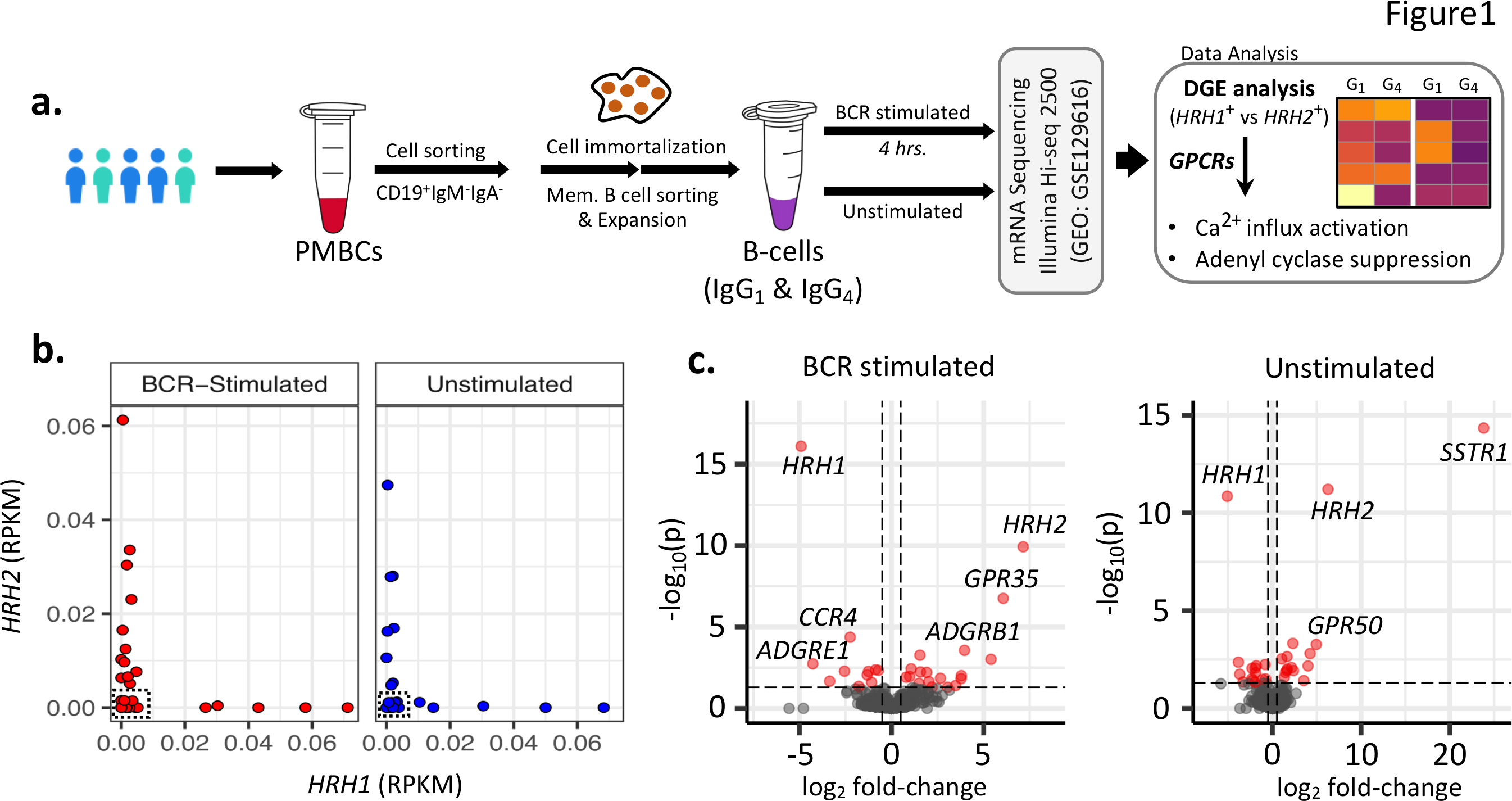
Title : Distinct and mutually exclusive Ca++ flux- and adenyl cyclase-inducing gene expression profiles of G-Protein-Coupled Receptors on human antigen-specific B cellsAuthors : Iris Chang1,2†, Abhinav Kaushik, PhD1,2†, Pattraporn Satitsuksanoa PhD1, Minglin Yang1, Laura Buergi Msc1, Stephan R. Schneider Msc1, Cezmi A. Akdis, MD1, Kari Nadeau MD, PhD2, Willem van de Veen, PhD1, Mübeccel Akdis, MD, PhD1*1 Swiss Institute of Allergy and Asthma Research (SIAF), University of Zürich, Davos, Switzerland.2 Sean N. Parker Center for Allergy and Asthma Research, Department of Medicine, Stanford University, Palo Alto, CA, USA.† Contributed equally* Corresponding authorB cells play an essential role in allergies by producing allergen-specific IgE, which is a prerequisite for allergen-induced degranulation of mast cells (MCs) and basophils. MCs, basophils, dendritic cells and bacteria are capable of releasing inflammatory mediators including histamine. Histamine is a bioactive amine that exerts its function through binding to histamine receptors (HRs), which are 7-transmembrane G-protein-coupled receptors (GPCRs). There are four types of HRs (HR1-4), wherein HR1 ligation triggers Ca2+ mobilization, HR2 stimulates and increases cAMP concentrations, and HR3 and HR4 inhibit cAMP accumulation1. In the presence of histamine in the environment, high affinity HR1is triggered causing cellular activation, followed by expression of 10 times lower affinity HR2 to regulate the over-inflammatory events. These HRs trigger different intracellular events upon activation, with HR1 as a Ca2+ flux-inducing activating receptor and HR2 as an adenyl cyclase-stimulating suppressive receptor 1,2. Therefore, to explore the response of B-cells in allergic diseases, we analyzed the expression profile of HRs and other GPCRs in B cell clones. We hypothesized that the expression profile of HRs (HR1+ vs HR2+ B cell clones) is associated with significant changes in the expression profile of other GPCRs that govern the downstream cascade of pathways associated with cAMP signaling or Ca2+ mobilization.A total of 27 IgG1 and IgG4 expressing B cell clones were isolated for gene expression analysis under BCR stimulated and unstimulated conditions (Figure 1A and Online Supplementary Methods) . Interestingly, we observed B-cell clones with mutually exclusive expression profile of HRH1 and HRH2 genes (Figure 1B), with more HRH1+ B-cell clones in BCR-stimulated samples than unstimulated samples. The subsequentHRH1+ vs HRH2+ differential gene expression analysis (Figure 1C ), reveal 27 differentially expressed (DE) GPCRs in unstimulated samples, with up-regulated P2RY13  and C5AR1genes  in HRH2 + B-cell clones (Figure 2A) , which are associated with the cAMP signaling and suppressive pathway3,4. To further prioritize the DE GPCRs specifically associated with Ca2+ and cAMP signaling pathways, we reconstructed the co-expression networks and performed the weighted degree analysis across HRH1+ vs HRH2+ clones. The analysis reveals that the purinergic receptor family of GPCRs (i.e. P2RY1 , P2RY13 )  and complement component 5a receptor family of genes (i.e. C5AR1  and C5AR2 ) share highest degree of interactions. These genes are up-regulated inHRH2+ samples and are well-known to affect cAMP signaling pathway3,4 (Figure S1A ). Intriguingly, we also observed upregulation of GPR35  in HRH2 + B cells, which is associated in maintaining a low baseline Ca2+ level5. Similarly, we also observed up-regulation of GPR68 and GPR171 in HRH1 + B cells; both are known to stimulate Ca2+ flux (Online Supplementary Discussion) .Similarly, 28 GPCRs were differentially expressed in BCR-stimulated samples (Figure 2B ), including higher expression of serotonin receptor type 1A (HTR1A ) and HCAR1  (or GPR81 ) inHRH2+ samples, with a cAMP-linked suppressive function. In addition, we also observed upregulation of complement component 5a receptor family of genes (i.e., C5AR1  and C5AR2 ) and GPR35 , in agreement with the trend observed in unstimulatedHRH2 + B-cell clones. Surprisingly, we observed a higher expression of prostaglandin E2 receptor subtype EP4 (PTGER4)  and adenosine A2A receptor (ADORA2A ) in HRH2+ samples3,6, which are known to be associated with activation of cAMP production and share the highest strength of interactions with the cAMP signaling sub-network (Figure S1B ). Among the up-regulated genes in HRH1 + samples, we found three Ca2+ mobilizing genes, i.e., GPR34 ,P2RY10  and PTAFR .The results reported in this study provides data for a novel hypothesis suggesting investigation of co-expressed genes that may play important synergistic or antagonistic regulatory roles in B-cell function.