Hanieh Radkhah

and 9 more

Background: Pulmonary thromboembolism (PTE) is a common complication of SARS-CoV-2, which raises the COVID-19 disease’s fatality rate from 3% to 45%. Nevertheless, due to fairly indistinguishable clinical symptoms and a lack of validated clinical prediction models, PTE diagnosis in COVID-19 patients is challenging. This study aims to investigate the applicability of hematological indices to predict PTE incidence and its severity in SARS-CoV-2 patients. Methods: A retrospective cohort study was conducted on hospitalized patients with a confirmed diagnosis of SARS-CoV-2 infection who underwent CT angiography to assess probable PTE in them. The correlation between CBC parameters one day prior to CT angiography and CT angiography outcomes, and simplified Pulmonary Embolism Severity Index (s-PESI) was investigated. Results: We discovered that among individuals with a probable PTE, males and those with higher platelet-to-lymphocyte (PLR) and neutrophil-to-lymphocyte (NLR) ratios had a greater likelihood of PTE incidence. PLR was a significant and independent predictor of PTE. Moreover, a higher neutrophil count was associated with a higher s-PESI score in COVID-19 patients developing PTE. Conclusions: Among haematological indices, NLR and more precisely PLR are cost-effective and simply calculable markers that can assist physicians in determining whether or not COVID-19 patients with clinically probable PTE require CT angiography and the higher neutrophil count can be employed as an indicator of PTE severity in COVID-19 patients. Further large multicenter and prospective studies are warranted to corroborate these observations.

Arash Pour Mohammad

and 4 more

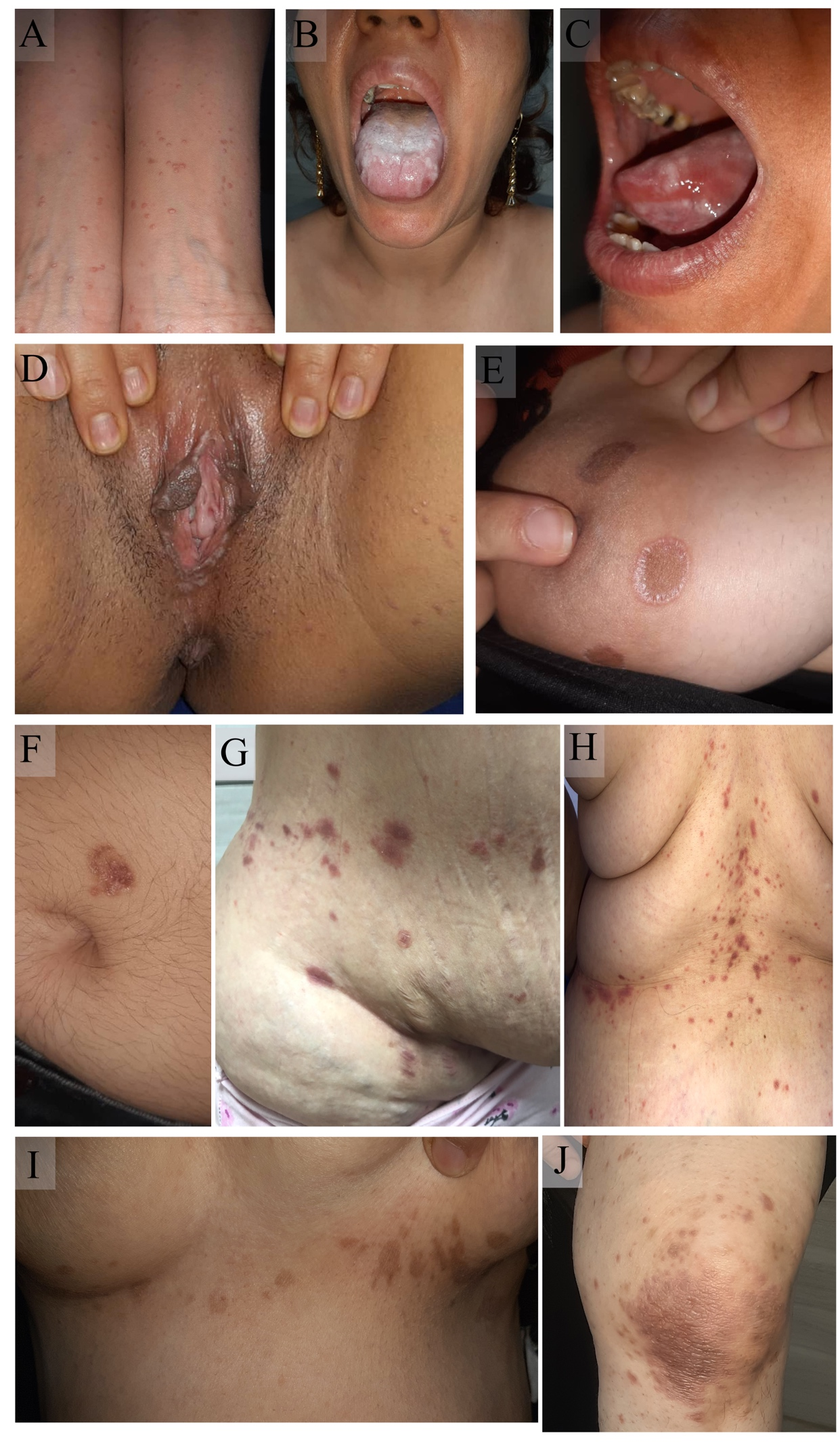
Mucocutaneous complications or adverse events due to SARS-CoV-2 infection or vaccination have been well-delineated in the literature, respectively. Most eruptions are considered to be mild and self-limiting; however, for the atypical cases which have a tentative clinical diagnosis, performing a biopsy and histopathological assessment is pivotal to confirm the diagnosis and subsequently prescribe a more tailored treatment. Despite the diverse reporting of such incidents globally, the rate of biopsied cases is restricted to less than 15% in most studies.    This case series elucidates 20 patients referred to the tertiary dermatology clinic, including 14 COVID-19 infection-related eruptions such as Lichen Planus (LP), Cutaneous vasculitis, Pityriasis rosea (PR), Discoid lupus erythematosus, Guttate psoriasis, Sarcoidosis, Raynaud’s phenomenon, non-specific lesions resembling genital warts, Beau’s line and one severe case of purpura fulminans with a promising outcome. Moreover, we presented 6 vaccine-induced cases comprising LP, Urticarial vasculitis, PR, Parapsoriasis, and Localized Morphea. The diagnosis of all cases has been proven by histopathological evaluation. We included pertaining anamnesis details of each patient together with vivid classifying images to pinpoint the morphologic features of each condition.   In line with our previous studies, the vaccine-induced eruptions were less severe compared to infection-related complications of COVID-19 and are mostly controllable by antihistamines and corticosteroids administration. Therefore, reporting such events should not hinder COVID-19 vaccination in the general population.