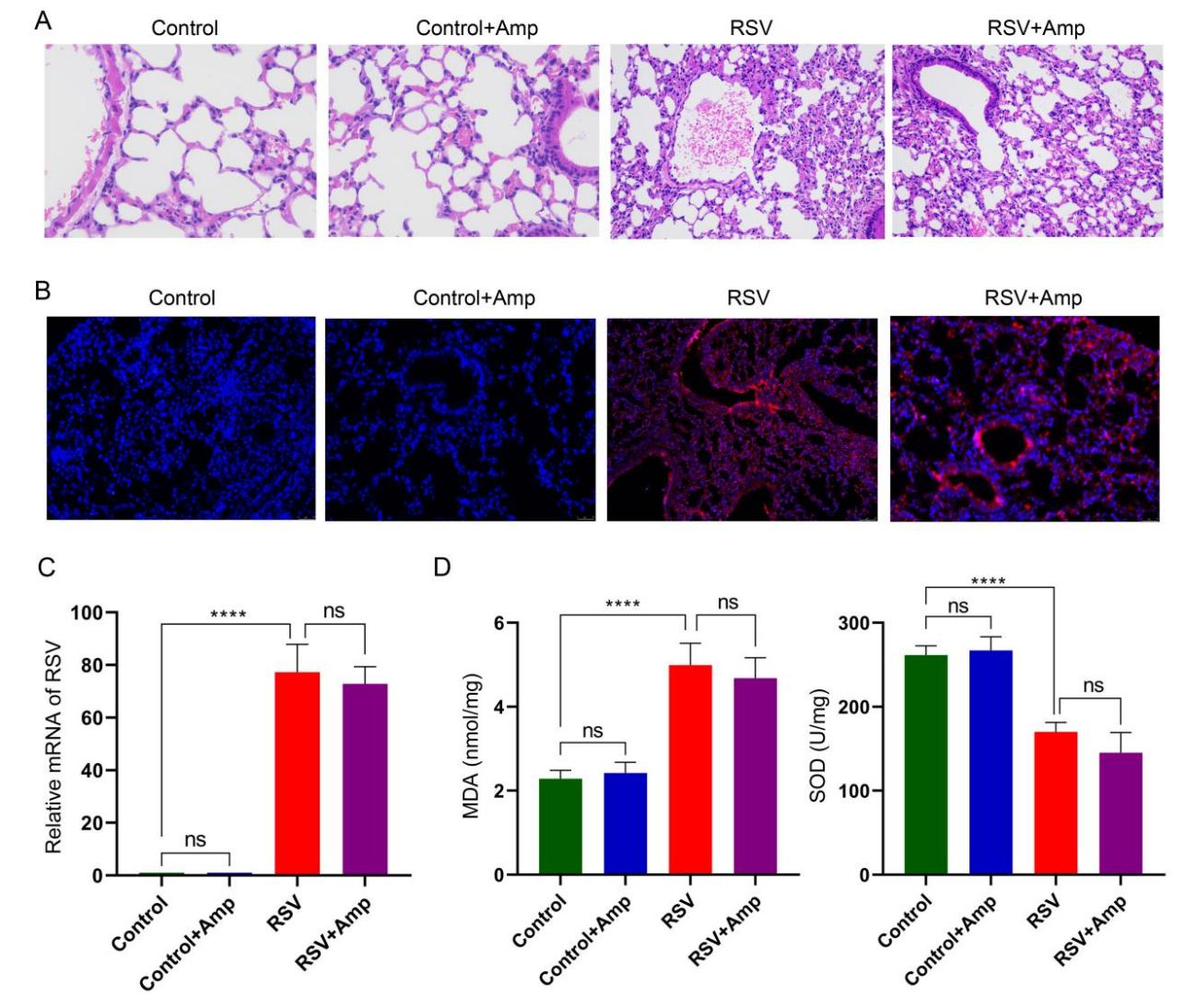
Background: The lung-brain-axis is an emerging biological pathway that is being investigated in relation to microbiome medicine. Increasing evidence suggest that pulmonary viral infections can lead to distinct pathological imprints in the brain, thereby the need to explore and understand this mechanism and find possible interventions. Objective: This study used RSV infection in mice as a model to establish the potential lung-brain axis phenomenon. We hypothesized that RSV infection could disrupt the lung microbiota, thereby compromising the immune barriers and thus induces significant shift in microglia phenotype. Methods: Mice were randomized into the Control, Ampicillin, RSV, and RSV+Ampicillin treated groups (n = 6 each). Ampicillin was given intratracheal instillation and seven days after the respective treatments, the mice were anesthetized. Hematoxylin-eosin (HE) staining of lung tissue to detect histopathology. Immunofluorescence label of specific target antigens in both the lung and brain tissues, namely, Malondialdehyde (MDA) and Superoxide dismutase (SOD) were used as markers of cellular damage. RT-qPCR was used to detect viral RNA in both tissues, ELISA to measure IL-1β, iNOS, IL-10 and Arg1 in the supernatant and 16s DNA technology were used to detect the lung microflora. Results: We found out that RSV infection induces elevated oxidative stress, reduced anti-oxidant and caused significant dysbacteriosis in the lungs of the mice. Pulmonary microbes were found affecting Th1-type immunoreactivity induced by RSV infection and eventually, microbiota in lung induced microglia phenotype shift in the brain of the mice. Conclusion. This study was able to establish that RSV infection can disrupt the pulmonary microbiome and immune barriers to induce microglia phenotype shift. Thus, we recommend a large sample size study with robust data analysis for the long-term effects of antibiotics and RSV infection on brain physiology.